
 Sign up with Google
 Sign in with Zoho

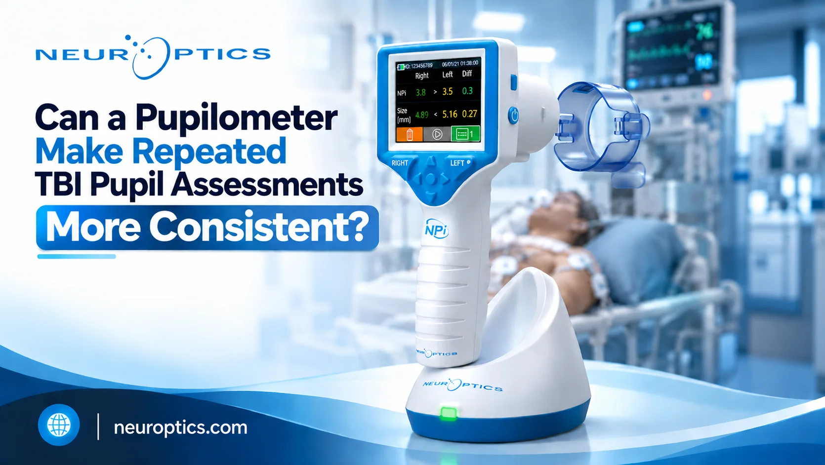
A pupil assessment performed at admission may look different from one completed several hours later. But how much of that difference reflects a change in the patient's condition, and how much could come from differences in observation or measurement?
For teams assessing Pupillary Response in Traumatic Brain Injury (TBI), consistency matters. Repeated examinations need to produce findings that clinicians can document, compare, and review. A quantitative pupilometer can support this process by measuring pupil size and reactivity objectively, rather than relying entirely on visual descriptions.
Why Can Repeated TBI Pupil Assessments Be Difficult to Compare?
Manual pupil assessments commonly use a penlight or flashlight to evaluate pupil size and response to light. Clinicians may describe the response as brisk, sluggish, or non-reactive. However, NeurOptics notes that these descriptions lack standardized clinical definitions and that manual assessments can be affected by examiner skill, visual acuity, pupil characteristics, and light positioning.
Its clinical overview reports that manual pupil assessment can involve up to 39% inter-examiner variability. This highlights a practical challenge: two assessments may not be directly comparable when observations depend on different examiners or subjective judgments.
For neurocritical care teams, the goal is not to eliminate clinical judgment. It is to make the pupil measurements being compared more objective and consistently documented.
What Does a Pupilometer Measure During a TBI Assessment?
A quantitative pupilometer measures characteristics of the pupil's response to a light stimulus. The NeurOptics NPi-300 provides objective measurements of pupil size, symmetry, and reactivity throughout the pupillary light reflex (PLR).
Its measurement set includes:
● Maximum and minimum pupil diameter
● Percentage change in pupil size
● Latency after the light stimulus
● Constriction velocity and maximum constriction velocity
● Dilation velocity
● Neurological Pupil Index (NPi®)
These measurements describe different aspects of the PLR. Rather than recording only whether a pupil appears reactive, clinicians can review quantitative information about the response. The results should still be interpreted within the broader neurological examination and the patient's clinical context.
How Does NPi® Add Context to Pupil Reactivity?
The NPi® is a numerical expression of pupil reactivity based on measured PLR variables compared with a normative model.
NeurOptics defines an NPi® from 3.0 to 4.9 as normal and a value below 3.0 as abnormal. An NPi® of 0 can indicate a non-reactive, immeasurable, or atypical response. These categories help clinicians describe measured reactivity using a standardized numerical scale.
For repeated TBI assessments, NPi® can be reviewed alongside pupil size and other clinical findings. It should not be treated as a standalone diagnosis or a replacement for the full neuro exam.
Why Baseline Measurements and Trends Matter
A single reading captures one moment. Repeated measurements create a record that clinicians can compare over time.
NeurOptics supports establishing baseline pupil measurements and reviewing subsequent results as trends. The NPi-300's SmartGuard stores up to 168 paired right and left pupil assessments for review during a patient's admission. This allows clinicians to look at previous measurements rather than relying only on the latest observation.
For teams monitoring TBI, a consistent process can include:
- Establishing a baseline measurement according to the unit's protocol.
- Repeating assessments at the prescribed intervals.
- Recording results consistently.
- Reviewing new measurements alongside previous findings.
- Following the clinical protocol when results change or require further assessment.
Trending helps organize the available data. Clinical interpretation and decisions remain the responsibility of qualified healthcare professionals.
How Can Hospitals Build a More Consistent Pupil Assessment Workflow?
Introducing a pupilometer is only one part of standardizing repeated assessments. Teams also need a clear process for using the device and reviewing its results.
NeurOptics identifies protocols, education, device availability, and documentation as important considerations when integrating quantitative pupillometry into clinical practice.
Hospitals can begin by defining who performs assessments, when measurements are taken, which values are documented, and how findings are communicated. Training should cover correct device positioning, obtaining a valid measurement, and following the manufacturer's Instructions for Use.
These steps help create a repeatable process across shifts and clinical teams. They also give hospital leaders a practical framework for evaluating how quantitative pupil measurement fits into existing neurological assessment workflows.
Conclusion: Make Repeated Assessments Easier to Compare
Repeated TBI pupil assessments are more useful when clinicians can compare findings collected through a consistent process. A quantitative pupilometer supports this goal by measuring pupil size and reactivity objectively and providing results that can be reviewed over time.
For hospitals and critical care teams, the value extends beyond a single reading. Combining standardized procedures, NPi®, baseline measurements, and trend review can support a more structured approach to assessing the Pupillary Response in Traumatic Brain Injury. Quantitative measurements complement the broader neuro exam; they do not replace clinical judgment.
FAQs
What does an abnormal pupil response mean after a TBI?
An abnormal finding requires clinical interpretation in context. Pupil measurements should be considered alongside the patient's overall neurological examination and established clinical protocols.
What is the difference between NPi® and the Glasgow Coma Scale (GCS)?
NPi® quantifies pupil reactivity, while GCS assesses levels of consciousness using eye, verbal, and motor responses. They describe different aspects of neurological assessment.
Can pupil assessment be performed when a patient is unconscious?
Pupil size and reactivity can be assessed in patients with impaired consciousness. The findings should be interpreted by trained clinicians as part of the overall neurological examination.
.webp)