 Sign up with Google
 Sign in with Zoho

.webp)
Most people don’t think about brain monitoring until they’re standing beside a hospital bed. It’s usually sudden. A fall from a staircase, a stroke during dinner, or a car accident that no one expected. And in that moment, everything narrows to one question:
Is there any indication of a brain injury?
When doctors walk in and perform a neuro exam, it can look deceptively simple. A light in the eyes, some commands or a quiet pause while they observe. For years, that assessment relied heavily on experience and visual judgment. Today, advanced neurological tools allow for something more precise.
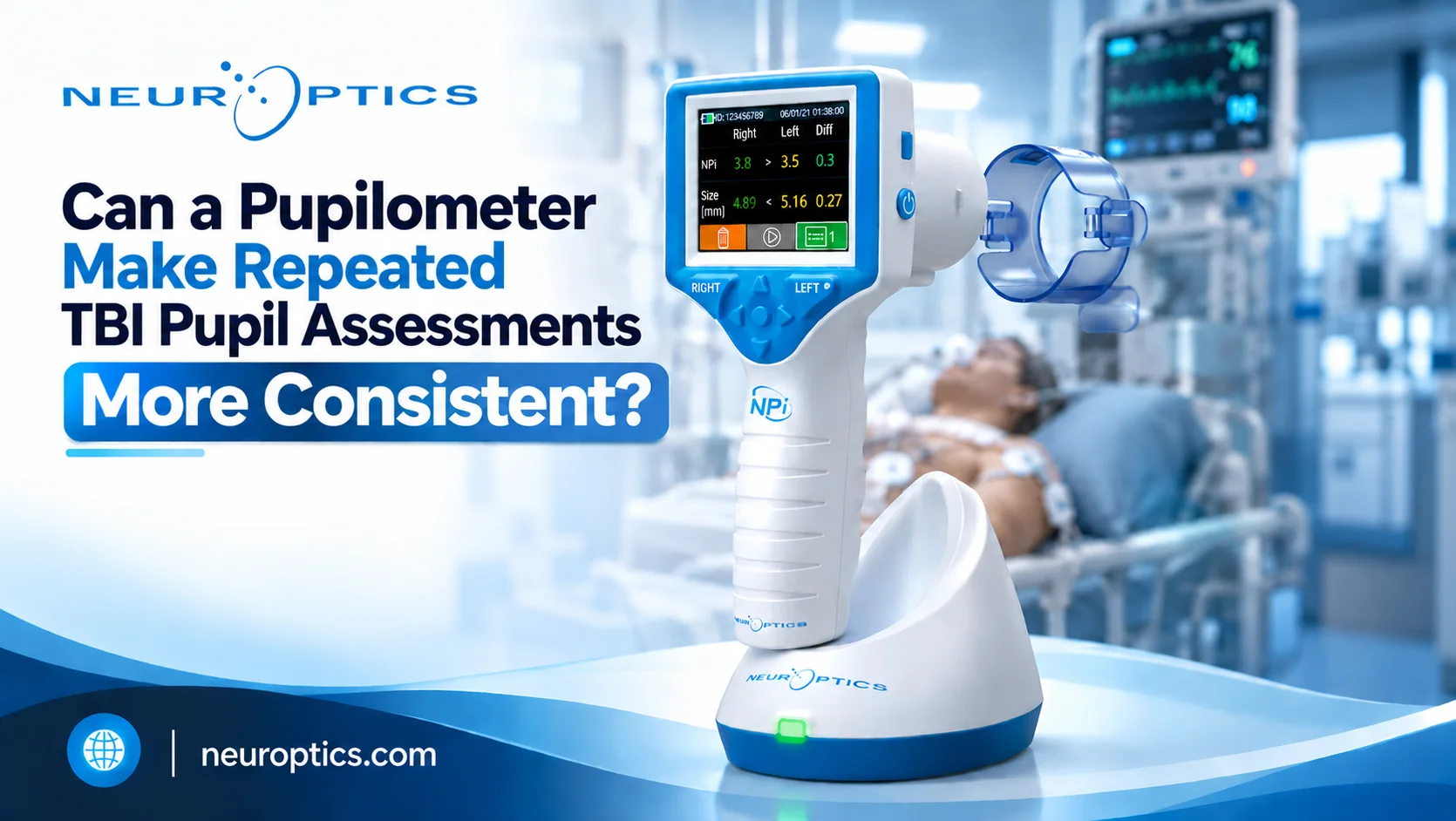
One of those tools is the Npi. Instead of describing a pupil as “slow” or “unequal,” clinicians can measure exact data points — including pupil dilation velocity and assign a standardized score. That shift from opinion to measurement has been studied carefully over the past decade.
Let’s walk through ten clinical findings that changed how this technology is used and why it matters beyond research journals.
Understanding the Clinical Parameters
Npi is one of the advanced neurological tools, which is a number between 0 and 5. It’s calculated through automated pupillary assessment and reflects how normally the pupil responds to light.
It evaluates:
- Size at rest
- Speed of constriction
- Reaction time
- Dilation speed
- Symmetry between both eyes
Generally, a score below 3 raises concern.
The important difference from the old penlight method? Two clinicians examining the same patient will get the same numeric output. That consistency is not a small improvement - in critical care, it’s significant.
Ten Foundational Clinical Studies Influencing NPi Implementation
1. Early Signals of Rising Brain Pressure
Several ICU-based studies observed that quantitative pupil assessment values often drop before measurable increases in intracranial pressure occur.
Brain swelling doesn’t always cause dramatic symptoms right away. It builds. Those early pupil changes can act as a warning system.
When detected in time, teams can:
- Adjust ventilation
- Administer osmotic therapy
- Prepare for surgical intervention
That kind of early move can prevent a crisis instead of reacting to one.
2. Traumatic Brain Injury Prognosis
Large multicenter studies involving TBI patients showed a clear pattern: persistently abnormal pupil reactivity scores were associated with poorer long-term neurological recovery.
What does that mean in real life?
It allows physicians to identify high-risk patients earlier. Families receive clearer expectations. Monitoring intensity can be adjusted based on measurable risk instead of guesswork.
It doesn’t predict the future with certainty, but it reduces ambiguity.
3. Stroke Deterioration Before Imaging Changes
In acute stroke units, subtle shifts in quantitative pupil assessment were sometimes observed before clinical decline was obvious.
In stroke care, minutes genuinely matter. Detecting worsening sooner can trigger repeat imaging and intervention before irreversible damage occurs.
The value here isn’t dramatic — it’s subtle and time-sensitive.
4. Cardiac Arrest Recovery Assessment
Neurological recovery after cardiac arrest is one of the hardest areas in critical care. Early exams can be clouded by sedation and metabolic factors.
Several studies found that abnormal quantitative pupil assessment readings within the first two days strongly correlated with poor neurological outcomes.
That data doesn’t replace compassion. It supports it. When families face difficult decisions, objective information matters.
5. Subarachnoid Hemorrhage and Vasospasm Prediction
Patients with subarachnoid hemorrhage face the risk of vasospasm days after the initial bleed.
Research showed that declining pupil reactivity scores often appeared before vasospasm became clinically obvious.
That allowed earlier treatment adjustments and reduced secondary brain injury in some cases. Preventing delayed complications improves rehabilitation outcomes significantly.
6. Sedation Management in ICU Settings
Sedation is necessary in many critical care patients, but balancing it is tricky. Too much delays recovery, too little can cause instability.
Studies showed that automated pupillary monitoring helped fine-tune sedation dosing.
The results were practical:
- Fewer ventilator days
- Reduced medication overuse
- Smoother neurological assessments
From a hospital operations standpoint, that translates into efficiency and lower complication costs.
7. Pediatric Neurocritical Care
Children cannot always describe what they feel. That makes neurological assessment even more complex.
Emerging data suggest that objective pupil measurement reduces uncertainty in pediatric intensive care settings.
It brings relief to parents knowing their child’s condition is being measured with precision rather than judged by sight alone. For clinicians, it provides solid, objective evidence they can rely on when making critical decisions.
8. Manual vs. Automated Assessment Comparison
Researchers have directly evaluated flashlight examinations against automated measurement methods in a widely discussed study.
The findings were straightforward:
|
Element |
Manual Exam |
Automated Measurement |
|
Subjective interpretation |
Yes |
Minimal |
|
Numeric scoring |
No |
Yes |
|
Trend tracking |
Limited |
Continuous |
|
Variation between providers |
High |
Low |
Reducing variability improves both safety and documentation clarity.
9. The Power of Trends Over Single Readings
One reading rarely tells the whole story. Several studies emphasized that gradual downward trends in quantitative pupil assessment were more predictive of deterioration than isolated low values.
In practice, that means clinicians can identify risk earlier — sometimes hours before visible neurological decline.
Trend data changes how urgency is communicated. It transforms “something feels off” into measurable evidence.
10. Integration Into Standard Protocols
Hospitals that incorporated Npi thresholds into stroke and TBI pathways reported smoother workflow and clearer escalation triggers.
Instead of debate, there’s structure.
When a defined number crosses a protocol threshold, the next step is clear. That reduces hesitation during emergencies.
Implications for Patients and Their Families
You may never operate these devices yourself. But if someone you love is hospitalized with a brain injury, this technology directly affects their care.
Understanding that objective monitoring exists can help you:
- Ask informed questions
- Understand clinical updates
- Feel more confident in the care plan
You don’t need to memorize scores. Just knowing assessment is measurable, not purely visual makes a difference.
The Operational and Financial Perspective
For healthcare leadership, implementation requires investment:
- Device acquisition
- Staff training
- Workflow adjustments
But long-term value often includes:
- Shorter ICU stays
- Reduced catastrophic deterioration
- Stronger documentation
- Lower legal exposure
Preventing even a small number of severe complications can offset initial costs.
Scalability is relatively straightforward once integrated into routine neuro exam protocols.
Frequently Asked Questions
Q: Does automated monitoring replace imaging?
A: No. It complements CT and MRI findings but does not replace them.
Q: Is automated pupillary assessment invasive?
A: No. It uses controlled light stimulation and is non-invasive.
Q: Does it guarantee better outcomes?
A: No single tool guarantees outcomes. However, earlier detection consistently supports earlier intervention, which improves overall care quality.
The Clinical Significance of Subtle Neurological Changes
Brain injuries rarely deteriorate without subtle warning signs. Those signs are often small. Slight asymmetry. Slower reaction. A gradual shift over hours.
Objective pupillary monitoring gives those small changes a measurable voice.
For families, that means fewer unknowns during frightening moments.
For clinicians, it strengthens decision-making.
For hospitals, it improves safety and operational clarity.
In neurological care, tiny changes can signal major shifts. Paying attention to them and measuring them accurately can truly change outcomes.
If your organization is reviewing neurological monitoring standards, take a close look at how pupil assessment is performed today. Integrating objective measurement into your neuro exam process doesn’t replace clinical expertise, it strengthens it where precision matters most.
Discover how Neuroptics can elevate your neurological assessment standards with precise, objective pupillary measurement technology.