 Sign up with Google
 Sign in with Zoho

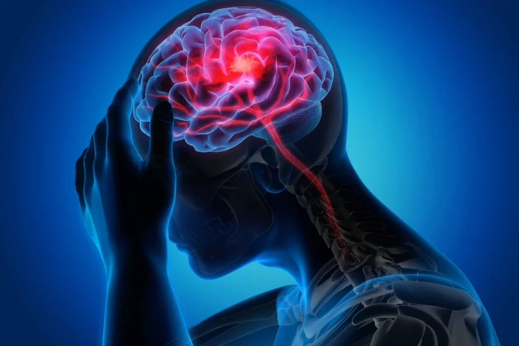
“This article details the clinical process doctors use to diagnose headaches at the base of the skull. It explores how physicians analyze symptoms, perform physical exams and use diagnostic nerve blocks to differentiate nerve irritation from muscle or neck issues.”
One of the most troublesome forms of pain is that which is located in the back of the head and has a pulsing, stabbing, or radiating quality to it. When a physician has to diagnose a case of occipital headache, they find themselves with an anatomical mystery. Indeed, (the occipital area) the part of the head situated at the lower back of the head and the upper neck is an area of enormous complexity, where vital nerves, large muscle groups and upper cervical vertebrae come together. Generally speaking, pain pathways in this area overlap in a major way, so what is felt as a mere strain may be a confused nerve or a referred pain from the spinal joint. And for patients, dealing with this uncomfortable feeling is an ordeal indeed.
Step 1: The Clinical Interview and Pain Mapping
Tests begin with a lengthy talk, not machine or needle. To map the headache's geography and behavior, doctors utilize pain mapping and precise inquiries because pain is subjective.
The exact nature of the sensation provides the first major clue to narrowing down the culprit:
- Electric Shocks vs. Steady Aches: Sudden, unilateral jabbing, scorching or electrical shocks from the neck to the scalp strongly suggest Occipital Neuralgia, a disorder with greater or lesser occipital nerve entrapment or irritation. A bilateral, dull, aching vice like grip is more like tension headache
- The Power of Triggers: Doctors will examine what causes or worsens pain. Does tilting the head or computer position hurt? Cervicogenic headaches (neck joint or ligament discomfort) are likely the cause. Does brushing your hair or sleeping on a pillow burn your skin? The classic sign of nerve irritation is allodynia
Ruling Out the Red Flags
Before diving deeper into benign headache types, clinicians must rule out rare but serious secondary causes using medical screening protocols. They screen for sudden onset thunderclap headaches, localized pain accompanied by a fever or stiff neck and any accompanying neurological deficits like vision changes, slurred speech, or loss of balance. Once these emergencies are off the table, the focused physical examination begins.
Step 2: The Physical and Neurological Exam
With a symptom map in mind, the doctor transitions to a hands on physical evaluation. The goal here is to physically reproduce the patient’s specific headache symptoms to confirm structural involvement.
Palpation and Tinel’s Sign
The physician will systematically press on specific anatomical landmarks at the base of the skull, directly over the pathways of the occipital nerves. In a healthy individual, this pressure might feel uncomfortable. However, in a patient with nerve related issues, pressing these suboccipital points will reproduce the exact shooting, radiating pain up into the scalp. This clinical phenomenon is akin to a positive Tinel’s sign, indicating hyper irritable nerve tissue.
Cervical Range of Motion and Sensory Testing
Next in the process is assessing the neck’s range of motion. Any limitation in motion, stiffness, or tenderness located in the area of the upper cervical spine (the C1, C2 and C3 vertebrae) gives support to the hypothesis of a cervicogenic origin. Lastly, basic neurological sensory tests are administered. Softly touching the head with a piece of cotton or a pin is performed to detect any areas of numbness, diminished sensation, or hypersensitivity thus helping the doctor identify what branches of the nerves are involved.
Step 3: Advanced Diagnostics Imaging vs. Nerve Blocks
When a physical exam points to a structural or neurological issue, advanced diagnostics are utilized. However, there is a common misconception that an MRI or CT scan will instantly provide a definitive headache diagnosis.
The Limits of Imaging
Magnetic Resonance Imaging and Computer Tomography scans are crucial for diagnosis. They help in diagnosis by ruling out the presence of conditions like herniated discs, severe osteoarthritis, vascular injury, or bone spurs that may lead to compression of the affected nerve root in the neck. However, the scans cannot be used for visualization of the inflammation or hypersensitivity inside the nerve system. As an instance, an individual may report severe nerve pain even if there is nothing abnormal in the MRI scan.
The Gold Standard: The Diagnostic Nerve Block
In order to obtain an accurate diagnosis of nerve related pain, physicians utilize an interventional process: the occipital nerve block.
If the patient experiences total relief, the diagnosis of occipital neuralgia is confirmed, allowing the medical team to implement long term nerve therapies. At this stage, a common concern for patients is understanding how long does occipital neuralgia last and whether the relief from these initial treatments will be permanent. If the pain remains completely unchanged, the doctor knows the root cause lies
If the patient feels completely relieved, then the diagnosis of occipital neuralgia is made. On the other hand, if the pain remains unchanged, the doctor will realize that the actual source is somewhere else.
Conclusion
This process of elimination involves wide patient history, highly localized physical examinations and targeted diagnostic injections to determine the reason for the base of skull discomfort. A careful clinical approach avoids patients from cycling through unsuccessful treatments since overlapping symptoms might mirror each other.
If you have pain in the area, keeping careful journal of triggers, pain trajectory and scalp response to touch will speed up next medical session and help your doctor solve the clinical riddle.
Disclaimer
This content is for informational purposes only and does not substitute for professional medical advice, diagnosis, or treatment. Always consult a qualified healthcare provider regarding a medical condition or symptoms at the base of your skull.