 Sign up with Google
 Sign in with Zoho


Cells have a sophisticated recycling system. When damaged organelles accumulate, misfolded proteins pile up, or nutrients run short, a process called autophagy is activated — engulfing cellular debris in double-membrane vesicles called autophagosomes and delivering them to lysosomes for degradation. ATG5 is one of the most critical proteins in this process, and researchers studying autophagy in almost any disease context will encounter the need to detect it. Tools targeting anti atg5 are foundational in autophagy research.
The Role of ATG5 in Autophagosome Formation
ATG5 (autophagy-related 5) is an E3-like ligase involved in conjugating ATG12 to anti ATG5 through a ubiquitin-like mechanism. This ATG12-ATG5 conjugate, in complex with ATG16L1, forms a large multimeric complex that localizes to the phagophore membrane during autophagosome biogenesis.
The primary function of this complex is to facilitate the lipidation of LC3 — the conversion of LC3-I to LC3-II — which anchors LC3 to the autophagosome membrane and is one of the most widely used markers of autophagy activation. Without ATG5, LC3 lipidation and autophagosome formation are severely impaired. ATG5 knockout models are therefore a standard tool for studying ATG5-dependent autophagy.
Why a Polyclonal Antibody for ATG5 Detection
Polyclonal antibodies contain a mixture of antibody molecules that recognize different epitopes on the same target. For ATG5, this breadth of recognition has practical advantages. The protein can undergo conformational changes and participate in multiprotein complexes that may partially mask individual epitopes. A polyclonal preparation is more likely to still bind the target even when some epitopes are obscured.
Polyclonal antibodies also tend to produce strong signals in tissue-based applications like immunohistochemistry, where signal intensity can be limited by the availability of secondary antibody binding sites. This is particularly relevant for ATG5, which is expressed at relatively low levels in many cell types under basal conditions.
Detection Formats and Their Applications
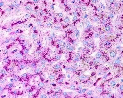
Immunohistochemistry (IHC): Localizing ATG5 in tissue sections allows researchers to determine which cell types are actively engaged in autophagy under different conditions — normal tissue, tumor samples, inflamed tissue, or tissue from disease models. ATG5 staining can be combined with other autophagy markers to build a more complete picture of the autophagic state.
Western blot: ATG5 migrates at approximately 55–60 kDa on SDS-PAGE, though it is often detected alongside its conjugated partner ATG12, appearing as a higher-molecular-weight band around 75–80 kDa. Observing the unconjugated versus conjugated forms provides information about the state of ATG12-ATG5 conjugation.
ELISA: Quantitative measurement of ATG5 in cell lysates or tissue homogenates allows comparison across large sample sets. This is particularly useful in drug screening studies where many conditions need to be compared simultaneously.
Disease and Research Contexts
Cancer biology: Autophagy has a complex relationship with cancer — it can suppress tumor initiation but support tumor cell survival under stress. ATG5 expression has been studied in relation to tumor progression, therapy resistance, and prognosis across multiple cancer types.
Neurodegeneration: In models of Parkinson's, Alzheimer's, and Huntington's disease, autophagy dysfunction contributes to the accumulation of toxic protein aggregates. ATG5 is a key marker for assessing whether the autophagic machinery is intact in affected neurons.
Infection and immunity: Many pathogens exploit or are targeted by autophagy. ATG5 plays a role in xenophagy — the autophagic elimination of intracellular pathogens — and in immune signaling pathways linked to innate immunity.
Metabolic stress: Starvation, hypoxia, and ER stress all induce autophagy. ATG5 expression changes in response to these stressors and is tracked as part of characterizing the cellular stress response.
Technical Notes for Researchers
ATG5 expression is relatively low at baseline and increases under conditions of nutrient deprivation or stress. Basal experiments should include a positive control — typically cells treated with rapamycin or subjected to starvation — to confirm that the antibody and protocol are performing correctly.
Tissue fixation method affects ATG5 detection in IHC. Formalin-fixed paraffin-embedded (FFPE) tissue often requires antigen retrieval with heat or enzymatic treatment to unmask the epitope. Frozen sections may give cleaner results but require more careful handling.
In Western blots, lysis buffer composition matters. ATG5 is a cytoplasmic protein, but its complex partners are membrane-associated during autophagosome formation. A lysis buffer that disrupts membrane interactions ensures complete recovery of both conjugated and unconjugated forms.
Conclusion
As autophagy research moves from characterizing the pathway's existence in various disease contexts toward understanding its precise regulation and therapeutic manipulation, the tools researchers use to detect key components like ATG5 need to keep pace. Selective autophagy — the targeted degradation of specific cargo like mitochondria (mitophagy), lipid droplets (lipophagy), or protein aggregates (aggrephagy) — is now an active frontier, and ATG5 remains central to many of these processes. Researchers entering this space for the first time, or those scaling from cell culture to in vivo models, will find that having well-validated, versatile ATG5 detection reagents is one of the foundational requirements for generating reliable data.